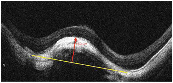
Fig. (9)
HD OCT scan showing a dome shaped macula, which is defined as inward bulge of RPE of more than 50 µm (red arrow), above a presumed tangential line (yellow line) joining the outer border of RPE, at the bottom of the posterior staphyloma.