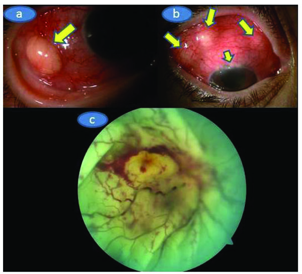
Fig. (1) a) Color picture of the temporal aspect of the right eye showing the significant hyperemia and whitish-yellowish subconjunctival mass (arrow), b) Multiple subconjuctival whitish yellowish masses and upper limbal multiple infiltrations (arrows), c) Color fundus picture of the right fundus depicting the massive exudative retinal detachment, palish optic disc with fuzzy borders, 360 degrees scattered retinal hemorrhages and whitish looking entire retina.