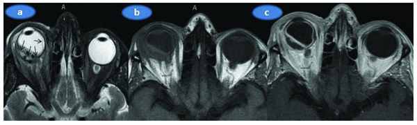
Fig. (2)
Orbital MR exam; a) The axial T2 weighted image; The right globe is deformed and the sclera is thickened especially in the posterior lateral aspect. The right retina looks thickened, undulating and detached (arrows) that is more prominent on the temporal side (arrows). The low T2 signal (relative to the vitreous) material (marked with asterix) has been deposited in both inside and outside of the detached retina implying the infiltrative nature of the deposition. Retrobulbar fat tissue, extraocular muscles and the optic nerve look unaffected. The eyelids on the right side look mildly edematous (thickened with a high signal), b) Axial T1 weighted image replicates the above-mentioned findings albeit less clearly. The subretinal collection has an intermediate signal intensity similar to the extraocular muscles, c) Axial postgadolinium T1 weighted image shows the thickening and contrast enhancement of the sclera and the retina while the subretinal collection did not exhibit enhancement.