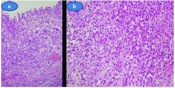
Fig. (3) a) A diffuse infiltrate of histiocytes with foamy or granular cytoplasm extending towards the lamina propria of the conjunctiva.Original magnificationX40, H and E stain. b) A few neutrophils, lymphocytes and plasma were also seen with diffuse infiltrates of histiocytes. Original magnificationX40, H and E stain.Three months after the initial presentation.