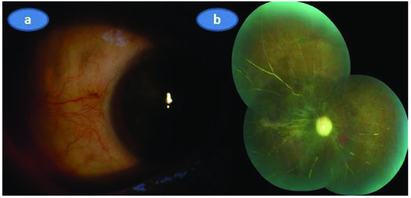
Fig. (4)
Three months after the initial presentation; a) Color picture of the right eye showing the temporal aspect of the right eye that was almost totally quitened, b) Composite color fundus picture of the right eye demonstrating the optic atrophy, ghost vessels and some retinal pigment epithelial changes related to severe infarct.