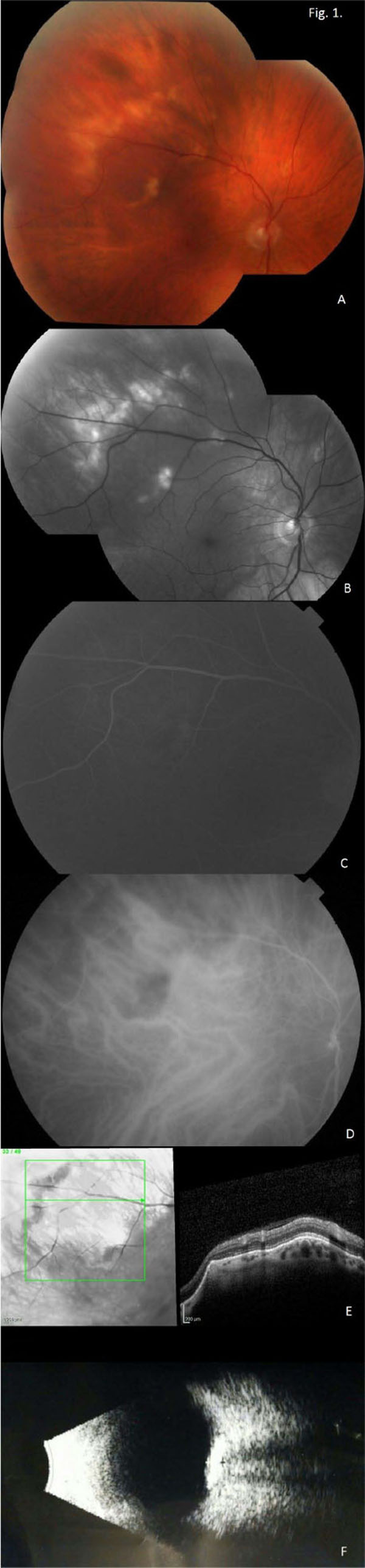
Fig. (1)
(Right Eye Images) A. Montage fundus photographs show yellow-white lesions in the superotemporal macula and periphery of the right and left eyes. B. Red-Free montage images highlight the lesions. C. Fluorescein angiogram shows staining. D. ICGA showed hyper-fluorescence in the corresponding region. E. OCT images show an elevated lesion of the choroid. F. B-scan ultrasound show solid appearing hyper-echoic elevations in the sclera and choroid with posterior shadowing.