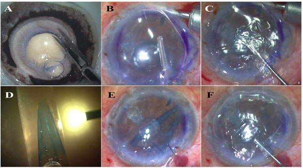
Fig. (1) A- Type I bubble dissected to form PDEK graft; B- Anterior Chamber Maintainer (ACM) connected to air pump is fixed and inferior peripheral PI performed with vitrector. Notice absence of bleeding because of air tamponade; C- Descemetorhexis performed under pressurized air infusion; D- E-PDEK technique used to enhance visualization of PDEK graft; E-Air infusion is stopped and air replaced with BSS. The PDEK graft injected into AC seen under microscope light; F- Graft unfolded and air injected to float it up. Notice small edge folds on the graft.