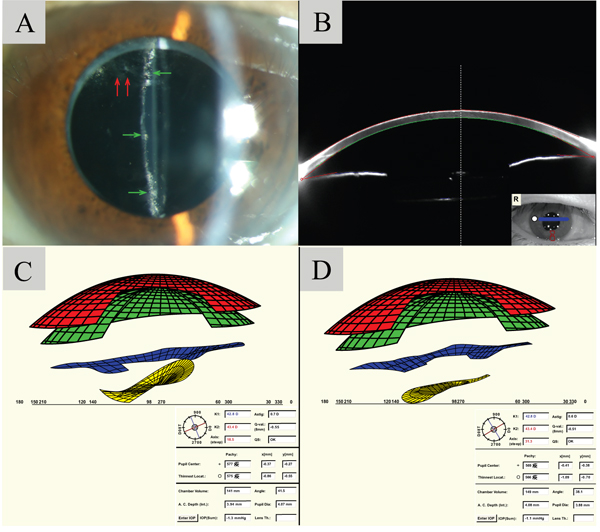
Fig. (2) A: Slit lamp examination reveals opacification and fibrosis of the posterior capsule (pointed by green arrows). Proliferating lens epithelial cells was noted on the superiorly within the pupillary zone (pointed by red arrows). B: Scheimpflug image before Nd:YAG laser. Mild accumulation of fluid is observed in the peri-IOL region, yet the contour of the IOL cannot be clearly visualized. C: Virtual eye simulation image before Nd:YAG laser capsulotomy was performed reveals a misalignment between the IOL (yellow) and the iris (blue). D: Virtual eye simulation showed that the iris (blue) and IOL (yellow) went back to normal alignment as soon as Nd:YAG laser capsulotomy was performed.