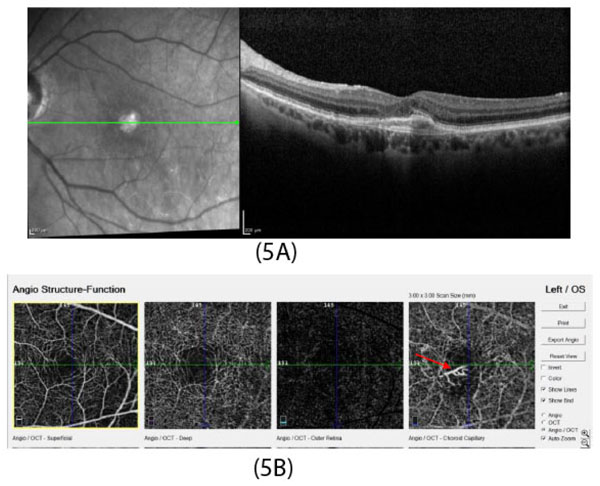
Fig. (5)
Multimodal imaging in a patient with type 2 CNV. OCT image (Fig. 5A) showing external limiting membrane (ELM) and ellipsoid zone (EZ) loss with subretinal hyper-reflective material.
OCT angiography image (Fig. 5B) shows type 2 CNV in choroidal capillary layer (red arrow).