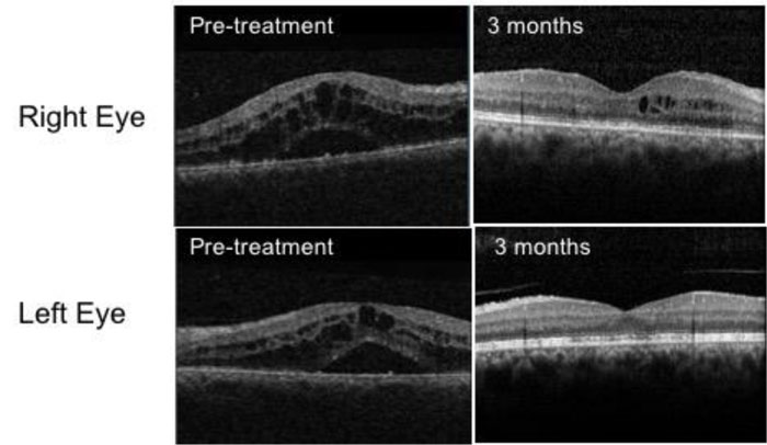
Fig. (1)
OCT images of Case 1 at pre-treatment and at 3 months. Pre-treatment images show gross Cystoid Macula Oedema (CMO) in both eyes with intra-retinal cysts and subretinal fluid. Improvement is seen at 3 months, corresponding to an improvement in visual acuity.