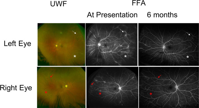
Fig. (2)
Case 2: Ultra-Wide Field (UWF) pseudo colour fundus photograph at presentation (OPTOS, California) and late phase wide field fundus fluorescein angiography at presentation and 6 months following treatment. Arrows and stars show corresponding areas of peripheral retinal vasculitis and sheathing of retinal vessels. FFA shows late leakage of fluorescein which resolved at 6 months. The lesions are asymmetric with the left eye more affected. Vitreous haze indicative of vitritis is seen in both UWF pictures.