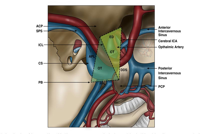
Fig. (4) Superior view of Cavernous Sinus (CS). The green area represents the Oculomotor Triangle (CT) and the yellow area represents the Carotid Triangle (CT). The dotted lines serve to represent the boundaries of the two triangles. The Interclinoid Ligament (ICL) divides the two triangles. The lateral border of the OT is limited by the Anterior Petroclinoid Ligament (APL) and the posterior border is limited by the Posterior Petroclinoid Ligament (PPL). The ICL forms the lateral border of the CT with the anterior borders and medial borders limited by the Endosteal Dura (ED) of the carotid canal and Dura of the Diaphragm Sella (DDS) medially. PCP = Posterior Clinoid Process, ACP = Anterior Clinoid Process, PB = Petrous Bone and SPS = Sphenoparietal Sinus.