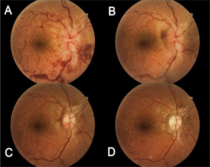
Fig. (1)
Fundus photography of the macula and optic nerve at initial presentation of the right eye (A) through the last visit (after 12 months) (D). The right eye demonstrated blurred margins of the disk with vascular tortuosity and flame-shaped hemorrhages and the macular area showed dot hemorrhages and granularity with peau d'orange at temporal midperiphery. (B) After 2 weeks, early resolution of papillophlebitis observed with less disk edema and hemorrhages; visual acuity dropped to 20/100. (C) Five-month color fundus photograph showed hyperemia of the disk and almost resolution of disk edema; angioid streaks were noted in the background. (D) At final presentation, after 12 months, no disk edema or hemorrhages were present; temporal pallor of the optic nerve was noted.

