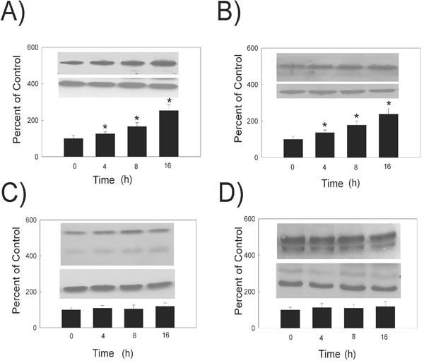
Fig. (3) Hydrogen peroxide treatment time dependence of PMCA protein levels. (Top Inset) A typical Western Blot showing time-dependent effects of 10 µM hydrogen peroxide on PMCA protein expression in the HLE B-3 cells. (A) PMCA1, 130 kDa (B) PMCA2, 120 kDa (C) PMCA3, 134 kDa (D) PMCA4 129 and 133 kDa. An equal amount of membrane protein was loaded in each lane and subjected to electrophoresis. Blots were incubated with a specific PMCA antibody, then stripped and reprobed with β-actin antibody antibody (lower Western Blot insert, 42 kDa). (Bars) Densitometric analysis of Western Blots. Results are presented as mean ± standard error of 3 or 4 separate experiments. A value of P ≤ 0.05 was considered significant (*).