Fig. (2)
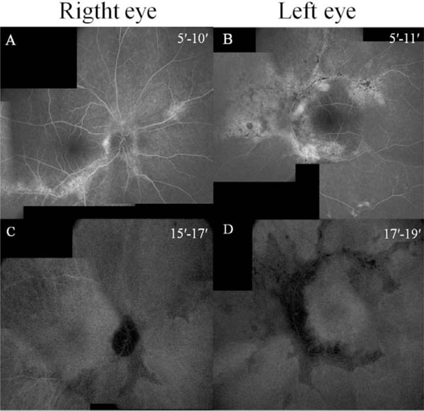
Fluorescein angiography shows a window defect (
A
,
B
) and indocyanine green angiography shows hypofluorescence (
C
,
D
) corresponding to the atrophy.