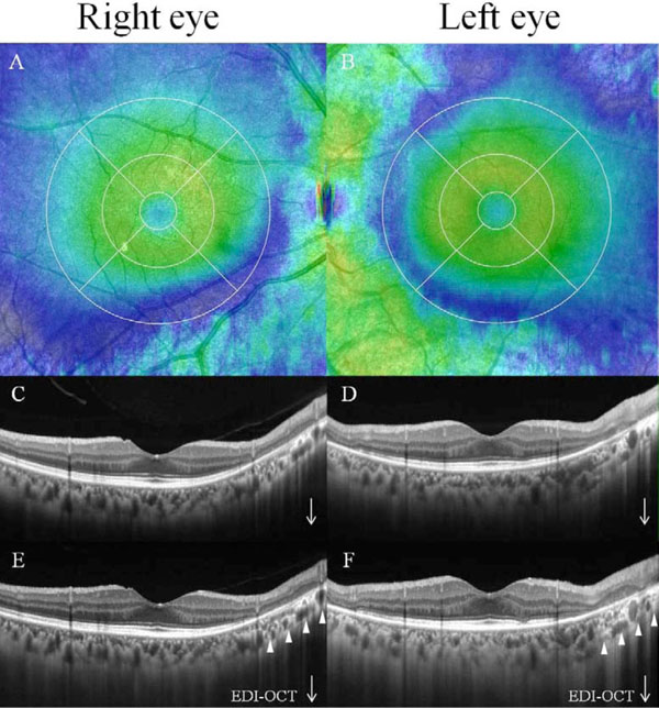
Fig. (3) Blue area of the color map reveals thinning of the retina on optical coherence tomography (OCT) in macular map mode (A, B).
Cross scans in the atrophic areas revealed thinning of not only RPE layer and the outer retinal layer, but also inner retinal layer (C, D).
Enhanced depth imaging OCT scans demonstrated that choroidal thickness was thin (arrowheads).