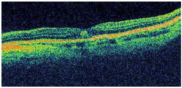
Fig. (2)
Optical coherence tomography of the left eye demonstrating a subfoveal lesion consistent with CNV.