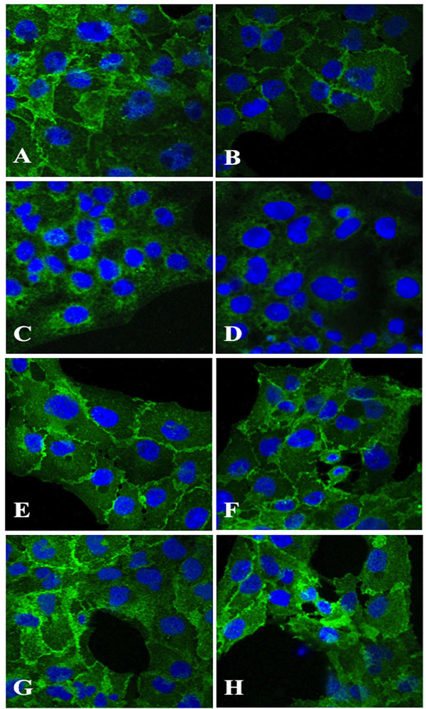
Fig. (3) Immunohistochemical staining of HLEC exposed 100 µM dexamethasone (B, D, F, H) for 24 hours indicated a decline in antigen
reactivity of N-cadherin (B) and α-catenin (D), whereas β-catenin (F) and γ-Catenin (H) showed no change in antigen expression as
compared to corresponding unexposed cells (A, C, E, G). Original magnification 1000x