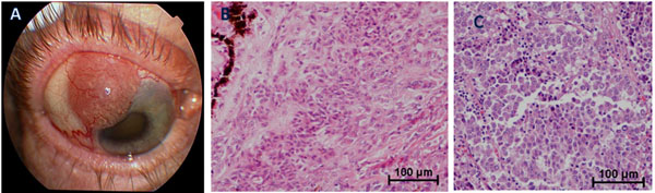
Fig. (1) (A) External photograph depicting extrascleral extension of amelanotic iris/ciliary body melanoma; (B) Histologic features of
primary tumor showing a mixed cell morphology with anaplastic epithelioid and spindle malignant melanoma cells invading the ciliary body.
Choroidal and iris involvement were also present (not shown); (C) Histologic features of metastatic liver lesion showing malignant
melanoma cells of epithelioid type with inflammatory infiltrate.