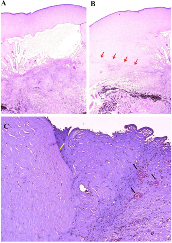
Fig. (2) Photomicrographs of the corneal surgical scar. (A) The anterior chamber angle was narrow with peripheral anterior synechiae, and a dense pupillary membrane. (B) Corneal scar tissue was noted circumferentially along the peripheral corneal margin (red arrow). (C) There were Bowman’s membrane fragments (yellow arrow) and thick, dense, fibrous tissue with inflammatory cells and small neovascular lumens (black arrow) present within the corneal scar. (hematoxylin & eosin, x25 (A & B), x100, (C)).