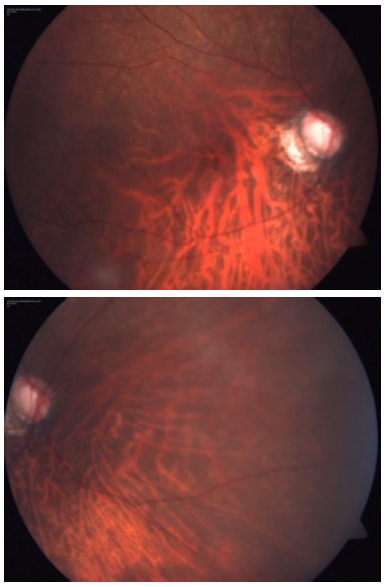
Fig. (3, 4) Fundus examination of both eyes under pupillary
midriasis revealed an oblique insertion of the optic disc, with a
myopic crescent, a posterior staphyloma, and it was also visible that
the optic nerve was cupping. There was mild macular atrophy on
both eyes and the peripheral fundus also presented important
atrophy.