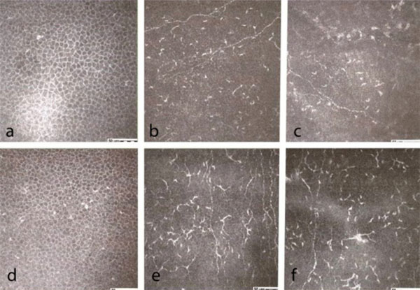
Fig. (4) Confocal microscopic evaluation in SSDE: a. intermediate epithelial cells, central corneal sectors; b. sub-basal nerve plexus, central
corneal sectors; c. sub-epithelial nerve plexus, central corneal sectors; d. intermediate epithelial cells, peripheral corneal sectors; e. sub-basal
nerve plexus, peripheral corneal sectors; f. sub-epithelial nerve plexus, peripheral corneal sectors.