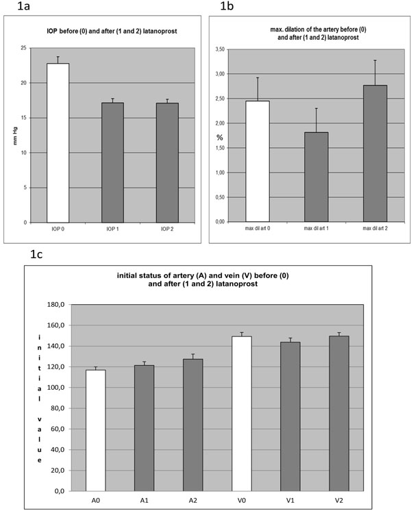
Fig. (1) (a) Intraocular pressure before (0) and after (1 and 2) beginning of the treatment with latanoprost; highly significant (p<0,01)
reduction of IOP by approximately 25%. Bars show mean +/- SEM. (b) Maximum dilation of the retinal artery (in %) as measured by DVA
after flicker stimulation before (0) and after (1 and 2) beginning of the treatment with latanoprost (+/- SEM); non-significant changes.
(c) Initial baseline value for the width of artery (A) and vein (V) given in measuring units (MU) assessed by DVA preceding flicker
stimulation before (0) and after (1 and 2) beginning of the treatment with latanoprost (+/- SEM); non-significant changes.