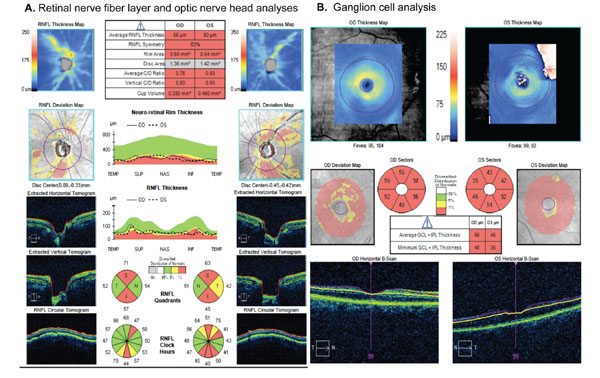
Fig. (2) Analyses of different scanning regions obtained with the Cirrus-OCT (Carl-Zeiss Meditec, Dublin, CA). A. Printout of retinal nerve fiber layer (RNFL) and optic nerve head analyses. There is diffuse loss of RNFL in both eyes, as indicated by the parameter average RNFL thickness. There is also neuroretinal rim thinning and enlarged cup, as indicated by the topographic parameters rim area, vertical C/D ratio and average C/D ratio. B. Ganglion cell analysis provided by the Cirrus-OCT indicating diffuse loss of the thickness of the combined ganglion cell and inner plexiform layers (GCL + IPL) in both eyes.