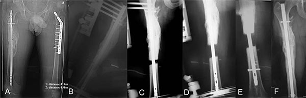
Fig. (1) (A) A 21-year-old man with 39mm post-traumatic shortening after intramedullary (IM) nailing of the right femur. Note successful bony union with no length discrepancy of the left femur. (B) Immediate post-operative anteroposterior (AP) x-ray showing the application of the hydrid external fixator parallel to the knee joint and to the nail. (C) Twenty days post-operatively AP x-ray showing progression of distraction. (D, E) Intraoperative x-ray after completion of the 39mm lengthening and insertion of locking screws and removal of the external fixator. (F) AP x-rays 6 weeks after insertion of interlocking screws. Note complete bony union.