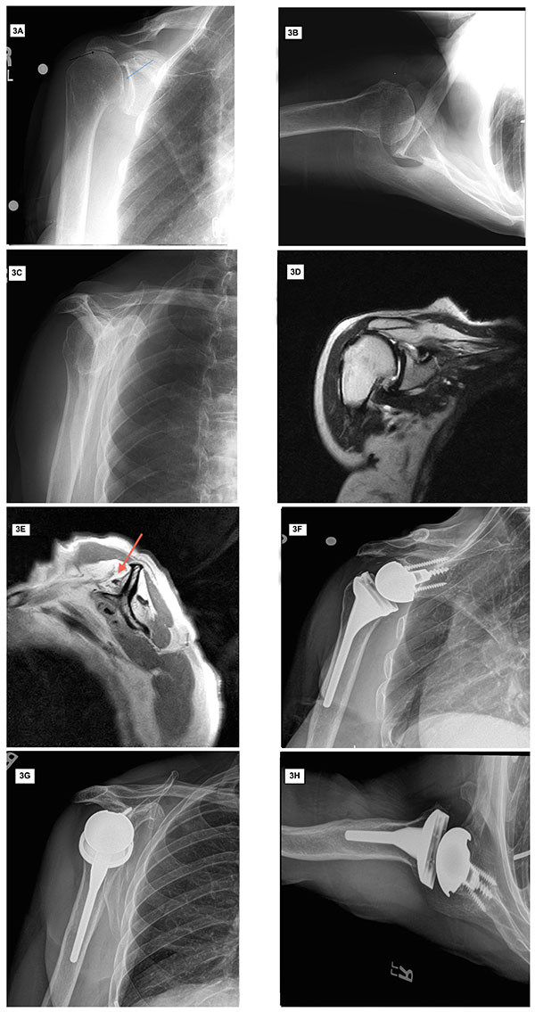
Fig 3 (A-H) Preoperative imaging and postoperative radiographs of patient with symptomatic irreparable rotator cuff tear with pseudoparalysis. Preoperative radiographs (A-C) demonstrate preserved glenohumeral joint space (blue arrow) and proximal migration of the humeral head with reduced acromiohumeral interval (black arrow). Preoperative T2 weighted coronal (D) and T1 weighted sagittal (E) MRI images demonstrate rotator cuff tear with advanced fatty infiltration and atrophy of rotator cuff muscle (red arrow). Postoperative anteroposterior and axillary views (F-H) demonstrate a reverse total shoulder arthroplasty.

