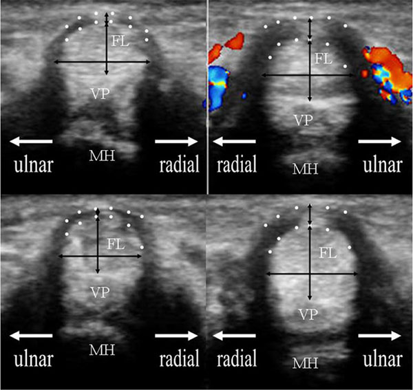
Fig. (2) The comparison of sonographic images between unaffected left ring finger (left image) and affected right ring finger (right image) of 69 years old woman including anatomical reference. The upper and lower images exhibit the transverse image at the MP joint in the neutral and hook grip positions, respectively. The flexor tendon was measured on the longitudinal and transverse axes: anterior–posterior thickness and radial-ulnar width. The hypoechogenic bundle including the A1 pulley at the rim of the flexor tendon, which is delineated by two dotted lines, was also measured on the top of the tendon. The A1 pulley thickness was defined as the longitudinal axis of this area, including the centralized hyperechogenic area. The black arrows indicate these measurements. The Dopplar flow around the A1 pulley was also recognized. FL: flexor tendon, VP: volar plate, MH: metacarpal head.