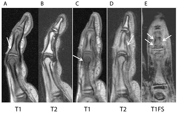
Fig. (2) T1- and T1-weighted fat-suppression magnetic resonance imaging (MRI) shows lesions with low signal intensity in the middle and proximal phalanges of the index finger (A, C, E). T2-weighted MRI showed high signal intensity in the intramedullary regions that showed low signal intensity on T1-weighted imaging (B, D).