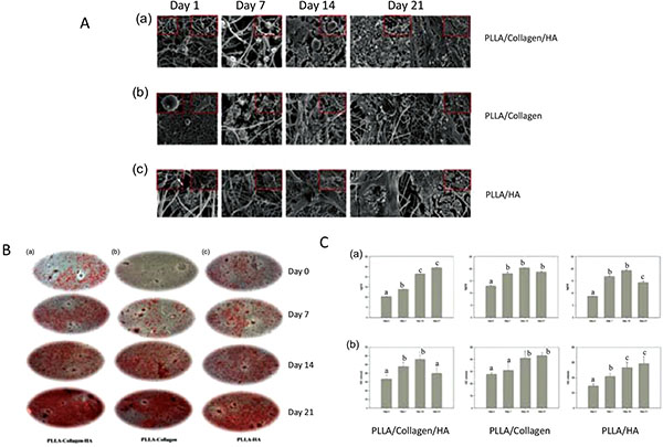
Fig. (1)
A Scanning electron microscopy images of (a) PLLA/Col/HA, (b) PLLA/Col and (c) PLLA/HA scaffold at different time points (0, 7, 14, and 21 days). B. Alizarin red images of the (a) PLLA/Col/HA, (b) PLLA/Col, (c) PLLA/HA scaffolds at different time points. C Levels of Osteocalcin (OC) and Calcium (Ca) in PLLA/Col/HA (a), PLLA/Col (b), and PLLA/HA (c) on Days 0, 7, 14, and 21. Abbreviations; PLLA; Poly(L-lactide), Col; Collagen, HA; Hydroxyapatite. Taken and reproduced with permission from [22].