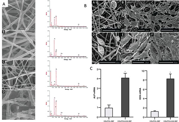
Fig. (2)
A The morphology and elemental composition of the four scaffolds.(A) COL/PCL with interconnected pores and smooth surface; (B) COL/PCL/nHA with a rough surface and a diameter of about 377 nm; (C) COL/PCL-SBF, few CaP deposits could be seen on the fiber surface; (D) COL/PCL/nHA-SBF, CaP precipitation was visible. B Scanning electron micrographs of PDLCs cultured on the COL/PCL-SBF and COL/PCL/nHA-SBF scaffolds. Cells on the COL/PCL-SBF (A, B, C) and COL/PCL/nHA-SBF (D, E, F) appeared to have no significant difference in morphology. The cells adhered to the fibers were spindle-shaped on the first day (A, D), and then extended gradually and adequately at day 3 (B, E). At day 8, the PDLCs were further flattened and stretched out flopodia. (C, F). C Real-time polymerase chain reaction analysis of ALP and OCN messenger ribonucleic acid expression in periodontal ligament cells on two scaffolds. Abbreviations: COL/PCL, the electrospun type-I collagen/poly(ε-caprolactone) scaffold; COL/PCL/nHA, the electrospun type-I collagen/poly(ε-caprolactone)/nanoscale hydroxyapatite scaffold; COL/PCL-SBF, the electrospun type-I collagen/poly(ε-caprolactone) scaffold immersed in simulated body fluid; COL/PCL/nHA-SBF, the electrospun type-I collagen/poly(ε-caprolactone)/nanoscale hydroxyapatite scaffold immersed in simulated body fluid; CaP, calcium and phosphorus compound. Taken and reproduced with permission from [23].