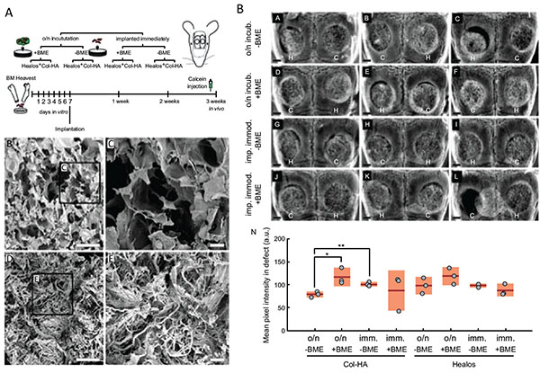
Fig. (3)
A Schematic of Experimental Design. Osteoprogenitors from the bone marrow were expanded in vitro before seeding to collagen-hydroxyapatite scaffolds and implanted in critical size calvarial defects. To label areas of active mineralization, calcein was injected intraperitoneally one-day prior to euthanization at three weeks post-implantation. (B) Electron micrograph of the Col-HA scaffold showing cellular morphology. Scale bar is 500 μm. (C) Enlarged inset from (A). Scale bar is 100 µm. (D) Electron micrograph of Healos scaffold. Scale bar is 500 µm. (E) Enlarged inset from (D). Scale bar is 100 µm. B Radiographs of cell-scaffold constructs after 3 weeks in vivo. (A)–(M) H and C denote Healos and Col-HA scaffolds, respectively. Scale bars are 1 mm. (N) Quantitation of radiographs. Light red bars indicate 95% confidence intervals and blue bars indicate one standard deviation. Taken and reproduced with permission from [35].