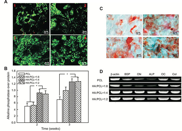
Fig. (3) Investigation into the in vitro effect of nHA nanoparticles on PCL: A) Cells seeded on PCL spiral scaffold (1), HA:PCL = 1:8 nano-HA/PCL spiral scaffold (2), HA:PCL = 1:4 nano-HA/PCL spiral scaffold (3), HA:PCL = 1:2 nano-HA/PCL spiral scaffold (4) for 7 days. Live cells were stained green, dead cells were stained red and nucleus were stained blue; B) ALP expression on the nano-HA/PCL spiral scaffolds (normalised against protein concentration) after 2 and 3 weeks in vitro. Data represent the mean ± standard deviation, n = 6. Significant difference between different material groups were denoted as * (p<0.05); C) Alizarin S Red staining of cells cultured on PCL spiral scaffold (1), HA:PCL = 1:8 nano-HA/PCL spiral scaffold (2), HA:PCL = 1:4 nano-HA/PCL spiral scaffold (3), HA:PCL = 1:2 nano-HA/PCL spiral scaffold (4) for 21 days; D) Gene expression of osteogenic markers in nano-HA/PCL spiral scaffolds analysed using representative electrophoresis gel after 3 weeks in culture [66].