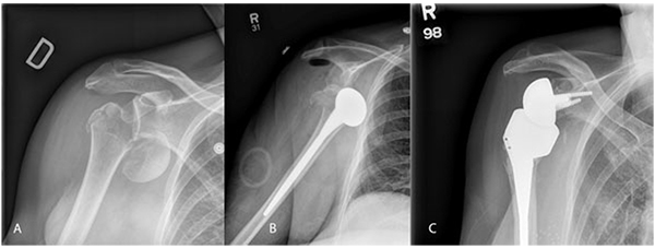
Fig. (1) A) plain xrays of comminuted acute proximal humerus fracture-dislocation. Note comminution of tuberosities and humeral head displacement with minimal calcar remaining. B) AP of post-operative day one humeral head replacement showing dislocation of prosthesis from glenoid. C) Six month post-operative follow-up AP imaging following revision to reverse total shoulder arthroplasty. Patient was doing functionally well with no complaints of pain and range of motion continuing to improve.