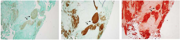
Fig. (5)
Histological examination of calcified deposits from Male Grade 3 Meniscus extracted after surgery. Serial sections were cut and stained. Left image, negative control. Middle image, stained with type X collagen. Right image, alizarin red stain. Arrows indicate an example of calcified deposits due to cellular hypertrophy. Images were taken with 4x objective.