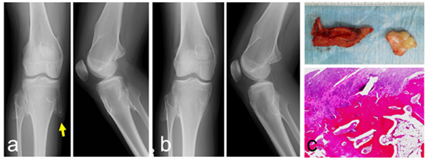
Fig. (4)
A 12-year-old girl with hereditary multiple exostoses. Plain radiographs show multiple protruding lesions arising from the distal femur, the proximal tibia and the proximal fibula (a). Radiographs after the lesion at the pes anserinus is resected (b) (left: antero-posterior view, right: lateral view). A photograph of a resected specimen (c: top). Histologically, hyaline cartilagninous cap and underlying lamellar bone trabeculae with fatty marrow are seen (c: bottom). (A yellow arrow indicates the tip of the osteochondroma causing the pain).