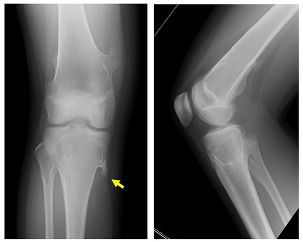
Fig. (5)
A 15-year-old girl with hereditary multiple exostoses. Plain radiographs show multiple protruding lesions arising from the distal femur, the proximal tibia and the proximal fibula (left: antero-posterior view, right: lateral view). (A yellow arrow indicates the tip of the osteochondroma causing the pain).