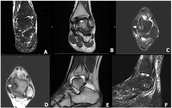
Fig. (2)
The MRI demonstrates the osteochondral lesions of the talus (OLT). The coronal plane of the MRI demonstrates anteromedial lesions of OLT, Hepple stage 1 and 2A in figure 2A, and 2B (arrow), respectively. The axial MRI demonstrates mid-medial lesions of the OLT, Hepple stage 2A in figure 2C and 2D. The sagittal MRI demonstrates non-displaced mid-medial lesion of OLT, Hepple stage 3, in figure 2E (arrow) and displaced mid-lateral lesion of the OLT, Hepple stage 4, in (Fig. 2F) (arrow).