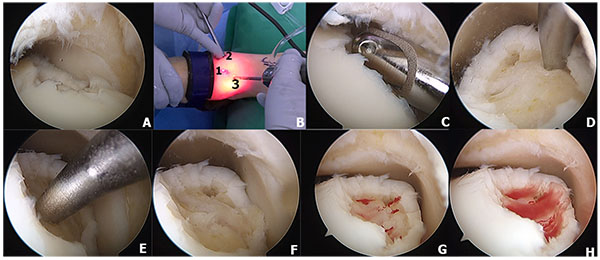
Fig. (3)
A microfracture technique of osteochondral lesion of the talus is demonstrated. A mid-medial osteochondral lesion of the talus is demonstrated on Fig. (3A). Anterolateral (2), anteromedial (1), and accessory portals (3) are demonstrated on Fig. (3B). The unstable cartilage rim was debrided using 4-mm shaver (3C). The chondral pick is used to create microfracture holes approximately 4-mm apart (3D-3E) and the base of the osteochondral lesion after complete debridement and microfracture (3F). After the tourniquet was deflated, the bleeding from the microfracture holes are demonstrated in Figure 3G-3H.