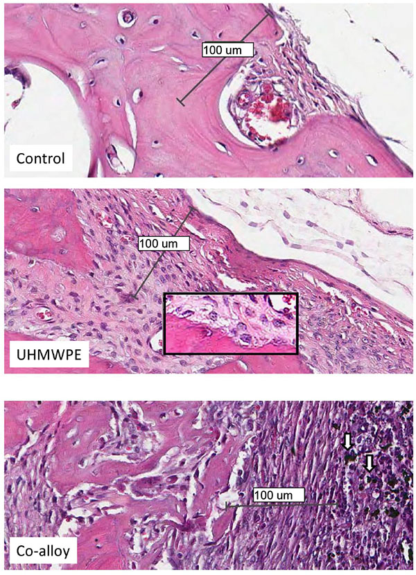
Fig. (5) Histological Analysis of Calvaria Tissue. Higher magnification of histologic sections of UHMWPE and Cobalt-alloy treated female calvaria (arrows indicate metal particles) show the lack of multinucleated osteoclasts at the bone pannus interface and instead show macrophage, fibroblast and osteoclast like cells associated with bone resorbtion eminating from the inflammatory pannus. There was a notable lack of lymphocyte infiltrates associated with both CoCrMo and UHMWPE particle inflamatory tissue. Neogenic woven bone was apparent in histological sections of all particle treated calvaria (Note:Bars indicate 0.1mm).