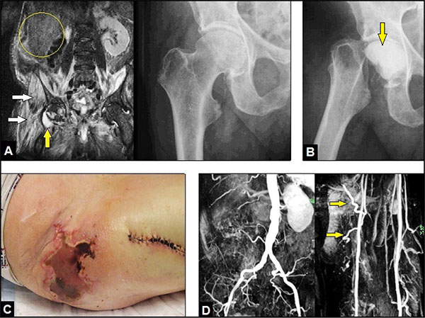
Fig. (1) : (A) Initial MRI demonstrating right IPM (white arrows) and fluid in right hip joint related to septic hip arthritis (yellow arrow) whereas in radiograph of the right hip there were no signs of infection-related osteolyses at this time. Note the formerly performed right nephrectomy (yellow circle). (B) Radiograph of the right hip showing performed GRA with the temporary insertion of a palacos cement spacer containing vancomycin (arrow) 1 week after first presentation. (C) Clinical photograph 2 weeks after first presentation showing right deep gluteal pressure-related soft tissue necroses due to MRSA infection. (D) MR angiography 4 months after first presentation demonstrating uneventful arterial blood flow through the aorta, both EIAs, both DFA's, and through both FCA's right (yellow arrows).