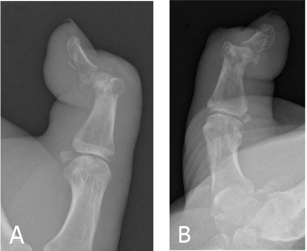
Fig. (1)
(a) Lateral and (b) anteroposterior radiographs of the left thumb showing a 4mm linear density anterior to the distal phalanx in addition to erosive/destructive changes in keeping with septic arthritis and adjacent osteomyelitis along with a pathological fracture of the neck of the proximal phalanx.