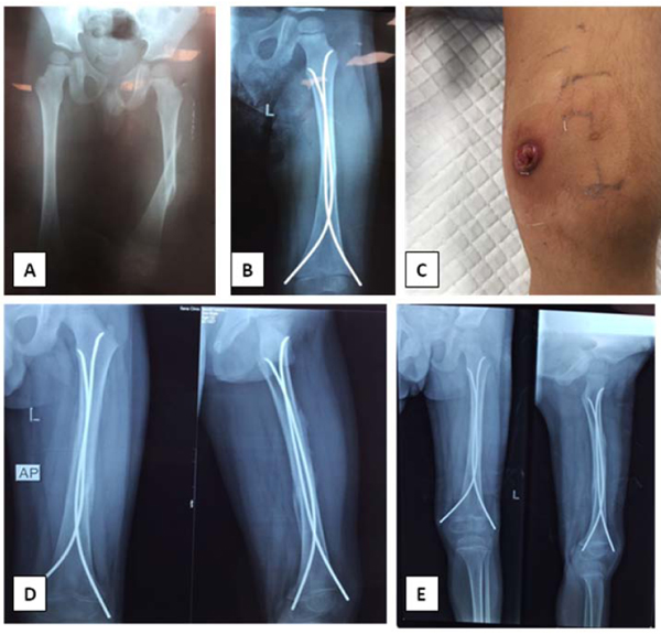
Fig. (3)
A 4.5-year-old boy who sustained a closed spiral fracture of the left femoral shaft due to a fall (low-energy trauma) and was treated with closed reduction internal fixation using flexible intramedullary nailing. (A) Preoperative radiograph. (B) Initial postoperative radiograph showing <50° varus. (C) Ulcer at the medial nail insertion site with superficial infection overlying the prominent nail. (D) Four-week follow-up showing a bridging callus at all cortices in both views. (E) Six-week follow-up showing that the fracture almost united; nails were removed 8 weeks after the first surgery, and the superficial infection healed.