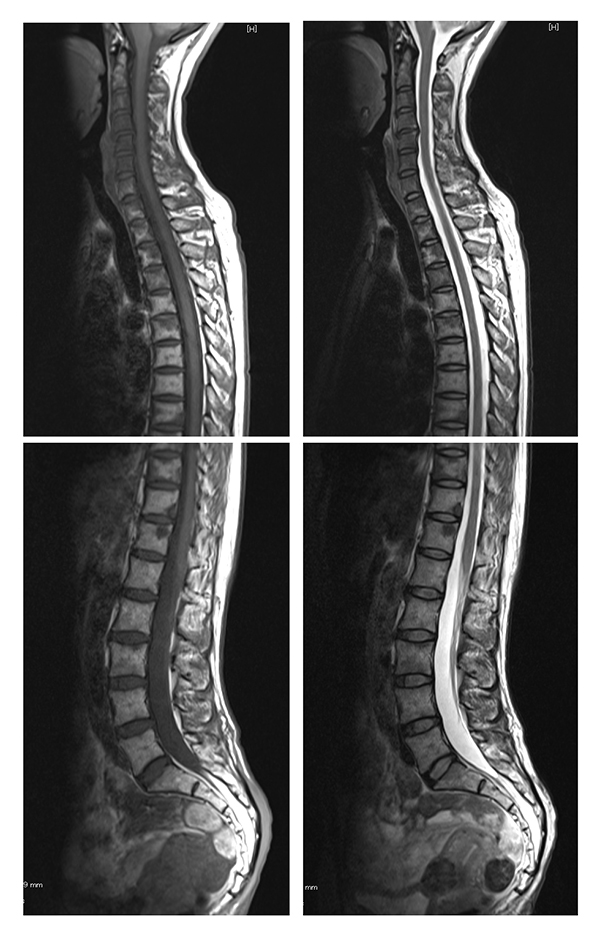
Fig. (3)
Bone marrow reconversion in a 47-year-old female. MRI demonstrates a diffuse lesion with low signal intensity in the whole spine on the T1 weighted image (left) and on the T2 weighted image (right). The signal intensity is slightly lower than that of the spinal cord on T1- and T2-weighted images.