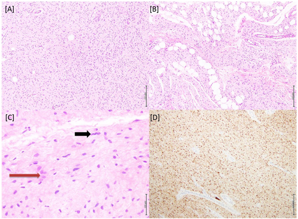
Fig. (1) [A] Microscopic picture of the benign spindle cell lesion. (H&E, 4X) [B] Another focus showing focal invasion into the fatty tissue included in the specimen. (H&E, 4X) [C] Diffusely arranged spindly (black arrow) to ovoid (red arrow) cells. (H&E, 10X) [D] The spindle cells showed strong positivity for S100 (4X).