Fig. (2)
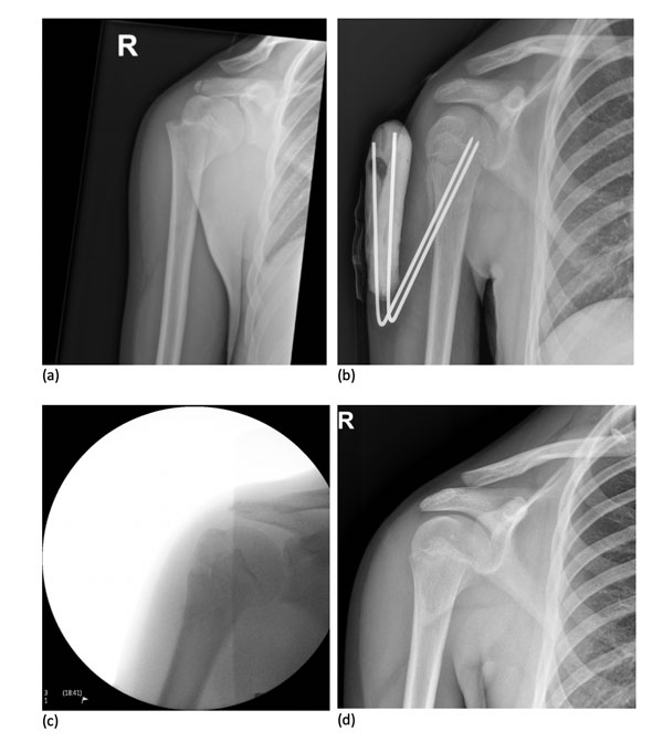
X-Ray images showing the progressive treatment using K-wires of a skeletally immature patient. Injury (
a
), K-wire insertion (
b
), outcome after removal (
c
), Radiograph at final follow-up (
d
).