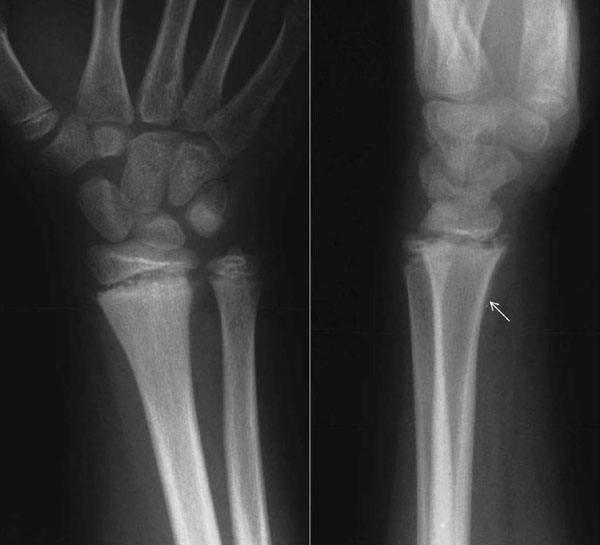
Fig. (1)
A 10-year-old boy forced his wrist into full extension while playing football. Clinical examination was indicative of a physeal fracture of the distal radius. The radiographs were negative. He was treated with a splint. Radiographs 5 weeks post-injury indicated a Salter-Harris type II lesion. The findings included widening of the distal radial physis on both views, widening between the palmar metaphyseal flake fragment and the metaphysis on the lateral view, and periosteal reaction on the palmar aspect of the radius (arrow).

