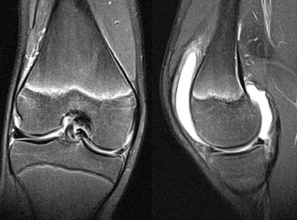
Fig. (4)
A 14-year-old boy sustained a rotational injury of the knee while playing football. Clinical examination was indicative of a physeal fracture of the distal femur. Radiographs were negative. A MRI was performed 6 weeks post-injury. The fat-suppressed images indicated knee effusion, widening and irregularity of the distal femoral growth plate demonstrating edema (high signal) along the physis, which indicates injury (coronal view) and uplifting of the periosteum from the posterior surface of the medial femoral condyle (arrow) with a small subperiosteal collection (sagittal view), most likely due to an occult Salter-Harris type I injury of the distal femur. No disruption and irregularity of the periosteum-perichondrium complex was noted.