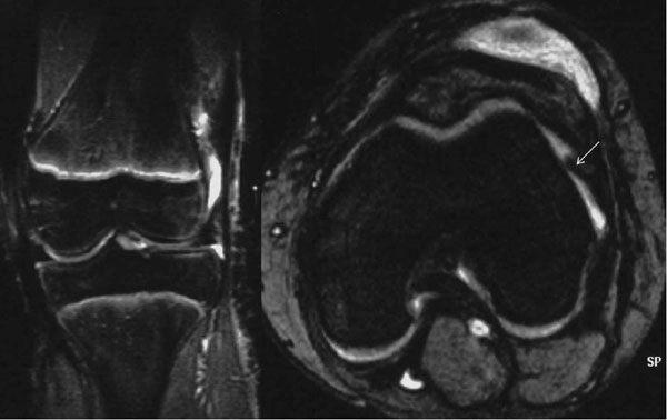
Fig. (5)
A 12-year-old girl fell while running with a bicycle. Clinical examination was indicative of a physeal fracture of the distal femur. Radiographs were negative. A MRI was performed 6 weeks post-injury. The fat-suppressed images indicated a prepatellar traumatic subcutaneous hematoma, widening and irregularity of the distal femoral growth plate demonstrating edema (high signal) along the physis, which indicates injury (coronal view) and local disruption of the periosteum (arrow) of the lateral femoral condyle (axial view), most likely due to an occult Salter-Harris type I injury of the distal femur.