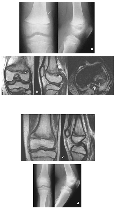
Fig. (6)
An 11-year-old boy injured his knee after a fall while playing football. Initial radiographs indicated no evidence of a fracture line. A clinical diagnosis of a potential Salter-Harris type I injury was made. Radiographs, 5 weeks post-injury, revealed widening of the distal femoral growth plate and periosteal reaction (arrow) along the distal lateral femoral metaphysis (a). A MRI was performed 6 weeks post-injury. Proton density (coronal and sagittal), as well as FL2D (axial) views, indicated a hypointense membrane interposed within the posterolateral portion of the disrupted physis, consistent with entrapped periosteum (arrows). There was bone bruising of the lateral femoral condyle (b). Proton density (coronal and sagittal) images also confirmed a medial metaphyseal Thurston-Holland fracture fragment with adjacent periosteal elevation (c). Radiographs, 2 years post-injury, showed incorporation of the periosteal reaction in the cortex and no altered growth (d). The final diagnosis was a Salter-Harris type II injury of the distal femur.