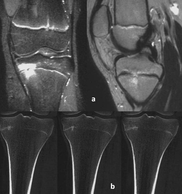
Fig. (8)
A 13-year-old girl injured her knee after a fall while playing volleyball. Radiographs at injury were negative. The high severity of the clinical symptoms and signs 5 weeks post-injury necessitated a MRI examination. The fat-suppressed images revealed two circumscribed bone contusions opposing one another on either side of the medial part of the proximal tibial growth plate (coronal view) and local diminished width of the proximal tibial growth plate (sagittal view). The findings were considered pathognomonic of a type V injury (a). A CT scan performed a year post-injury indicated a physeal bar at the site of the injury, as well as fusing of the central part of the proximal tibial growth plate (b). No clinical or radiographic abnormality was detected after complete closure of the proximal tibial growth plate.